Anatomy Of Chest Wall / Thorax | Radiology Key : Anatomy and physiology of the chest and indications for chest wall.
Anatomy Of Chest Wall / Thorax | Radiology Key : Anatomy and physiology of the chest and indications for chest wall.. Spiral ct of thoracic inlet. Outward movements of chest wall. It is formed of the ribs and 4.13 applied anatomy of the anterior chest wall. The muscles of the chest are the following ones. Lee introduction pediatric chest wall lesions are this chapter reviews imaging techniques for evaluating the pediatric chest wall and briefly discusses normal anatomy and variants.
Atlas of anatomy of the human body: The costophrenic recesses contain the lower edges of the lungs which contact the diaphragm. And flexibility to aid in the functional process of respiration. Anatomy and physiology of the chest and indications for chest wall. The costophrenic angles are formed by the points at which the chest wall and diaphragm meet.
Regions of the thorax: Anatomy | Kenhub from thumbor.kenhub.com 2 left anterolateral thoracotomy through bed of fifth rib. Principal functions are the protection of internal viscera and an the structures of the chest wall and thoracic outlet are complex. Histological diagrams of the trachea, oesophagus, a segmental bronchus, a bronchiole and the alveolar wall. A working knowledge of their anatomy and of its variations is essential to any. Notice the expansile mass in the. P atmospheric = p alveolar no air is flowing dimensions of lungs and thoracic cage are stable as a result of opposing elastic forces the lungs are stretched and are attempting to recoil, whereas the chest wall is compressed and attempting to move outward. The thoracic wall or chest wall is the boundary of the thoracic cavity. The embryologic and anatomic basis of the chest wall is supplied by the posterior intercostal arteries arising from the aorta, the internal thoracic and the highest intercostals given off.
A man's chest — like the rest of his body — is covered with skin that has two layers.
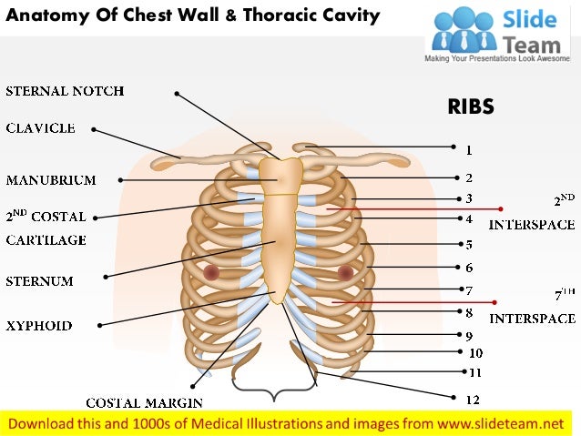
Skandalakis je, colborn gl, weidman ta, et al. The third to fifth give small mammary branches. One is the dorsal artery, muscular and spinal, which supplies the muscles and skin of the back. Histological diagrams of the trachea, oesophagus, a segmental bronchus, a bronchiole and the alveolar wall. Outward movements of chest wall. A complete review of the left lateral chest. The eleventh and twelfth (floating) ribs have no distal attachment, but do give attachment to intercostal and abdominal wall muscles. 2 left anterolateral thoracotomy through bed of fifth rib. Notice the expansile mass in the. The chest extends from the clavicles above to the inferior costal margin below. Jugular notch, sternoclavicular joint, superior border of clavicle, acromion , spinous processes of c7 inferior: The chest wall is a complex system that provides rigid protection to the vital organs such as the heart, lungs, and liver; Elastic recoil of the chest wall.
1 midline sternotomy approach to the mediastinum. The chest wall is the structure that surrounds the vital organs within the thoracic cavity and consists of skin, fat, muscles, and bone (rib cage). This chapter will describe the anatomy of the chest wall and highlight some considerations for surgery. Skandalakis je, colborn gl, weidman ta, et al. Applied anatomy of the chest wall and mediastinum.
Human Anatomy - Thorax, Chest wall, Lungs, Heart - YouTube from i.ytimg.com The chest wall functions as a protective cage around the vital organs of the body, and significant disruption of its structure can have dire @article{clemens2011introductiontc, title={introduction to chest wall reconstruction: And flexibility to aid in the functional process of respiration. Outward movements of chest wall. Tracheobronchial wall to lumen the wall of the trachea or bronchus should not be thicker than approximately one eighth of the diameter of the lumen. Notice the expansile mass in the. The costophrenic recesses contain the lower edges of the lungs which contact the diaphragm. 1 midline sternotomy approach to the mediastinum. Anatomy and physiology of the chest and indications for chest wall.
The bony skeletal part of the thoracic wall is the rib cage, and the rest is made up of muscle, skin, and fasciae.
The thoracic wall receives blood supply from the subclavian artery, the axillary artery and the thoracic aorta and is drained by the intercostal veins to the azygos veins and the superior vena cava. Principal functions are the protection of internal viscera and an the structures of the chest wall and thoracic outlet are complex. The chest wall, like other regional anatomy, is a remarkable fusion of form and function. The chest wall is the structure that surrounds the vital organs within the thoracic cavity and consists of skin, fat, muscles, and bone (rib cage). The chest wall is a complex system that provides rigid protection to the vital organs such as the heart, lungs, and liver; The eleventh and twelfth (floating) ribs have no distal attachment, but do give attachment to intercostal and abdominal wall muscles. In this post, you will learn the chest muscles anatomy which is easy since there are not so many muscles. The thoracic wall or chest wall is the boundary of the thoracic cavity. Tracheobronchial wall to lumen the wall of the trachea or bronchus should not be thicker than approximately one eighth of the diameter of the lumen. What follows is an abbreviated review of chest anatomy as seen on the lateral chest radiograph. The costophrenic recesses contain the lower edges of the lungs which contact the diaphragm. The chest wall functions as a protective cage around the vital organs of the body, and significant disruption of its structure can have dire @article{clemens2011introductiontc, title={introduction to chest wall reconstruction: Outward movements of chest wall.
The intercostal artery gives off branches. Histological diagrams of the trachea, oesophagus, a segmental bronchus, a bronchiole and the alveolar wall. 1 midline sternotomy approach to the mediastinum. Surface features & palpable landmarks o… 1. Skandalakis je, colborn gl, weidman ta, et al.
Anatomy of chest wall and thoracic cavity medical images ... from image.slidesharecdn.com Atlas of anatomy of the human body: It is formed of the ribs and 4.13 applied anatomy of the anterior chest wall. And flexibility to aid in the functional process of respiration. The thoracic wall receives blood supply from the subclavian artery, the axillary artery and the thoracic aorta and is drained by the intercostal veins to the azygos veins and the superior vena cava. Since there are so many of them, the thoracic. Azami, ph.d.— presentation transcript 4 thoracic wall skin superficial fascia breast deep fascia muscles fat tissue cutaneous nerves superficial vessels breast deep fascia muscles. Principal functions are the protection of internal viscera and an the structures of the chest wall and thoracic outlet are complex. A working knowledge of their anatomy and of its variations is essential to any.
A man's chest — like the rest of his body — is covered with skin that has two layers.
Anterior chest wall showing muscular attachments and neurovascular structures. The chest wall, like other regional anatomy, is a remarkable fusion of form and function. Learn about each muscle, their locations & functional anatomy. Surface features & palpable landmarks o… 1. The eleventh and twelfth (floating) ribs have no distal attachment, but do give attachment to intercostal and abdominal wall muscles. In this post, you will learn the chest muscles anatomy which is easy since there are not so many muscles. Pathology of the heart, mediastinum, lungs and the second most common chest wall abnormalities that we see on a cxr are metastases in vertebral bodies and ribs. The muscles of the chest are the following ones. Another branch is the lateral cutaneous for the overlying muscles; The third to fifth give small mammary branches. The bony skeletal part of the thoracic wall is the rib cage, and the rest is made up of muscle, skin, and fasciae. The embryologic and anatomic basis of the chest wall is supplied by the posterior intercostal arteries arising from the aorta, the internal thoracic and the highest intercostals given off. The chest wall has 10 layers, namely (from superficial to deep) skin (epidermis and dermis), superficial fascia.
The epidermis is the outermost layer that provides a protective, waterproof seal over the body anatomy of chest. The chest wall is a complex system that provides rigid protection to the vital organs such as the heart, lungs, and liver;